
Sipping espresso in the café, I hear the background noise of the espresso grinder mingling with the murmur of voices. The couple next to me are chatting in Japanese, drinking coffee and downing a pastry. I don’t understand a word, but still I can hear that each person’s larynx is functioning well.
The larynx is a valve, designed to control the flow of food, air and sound. Each person takes a sip of coffee, swallows, breathes in and then engages in conversation. Liquids and solids are separated from the air each individual breathes, then directed down the esophagus. Air is directed between the vocal cords into his and her lungs. The air is put to use again to make sound on its way back out. The larynx does all of this work, all very rapidly and all rather precisely.
Thus, the larynx is basically a talented valve, with three main functions: regulate breathing, create sound, and keep food and liquids out of the lungs. Of course, we have all seen someone blast his friend with drink when he rushes his larynx and tries to swallow and talk simultaneously. It helps to have a microsecond or two between functions.
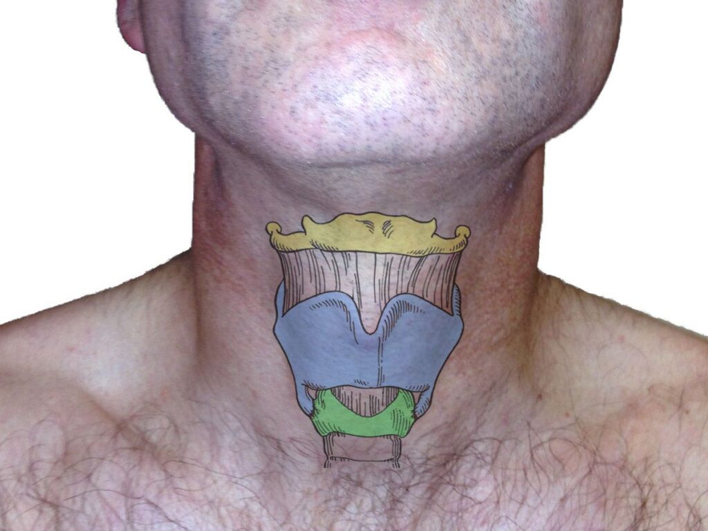
The Cartilages
As the young man beside me swallows his coffee, I see the bulge of his “Adam’s apple” — or medically speaking, the thyroid cartilage — moving up and down in his neck. The soft thyroid cartilage enlarges when exposed to testosterone, perhaps partly for evolutionary mating reasons. The net external effect is that it protrudes visibly. On the inside, testosterone thickens and elongates the vocal cords. The longer and thicker they end up, the lower the notes they are capable of producing. The thyroid cartilage sits above and articulates with the cricoid cartilage. Below the larynx is the windpipe or trachea, which you can feel in some people with thin necks. Above the Adam’s Apple, the hyoid bone helps suspend the larynx in the neck.
How We Look Inside
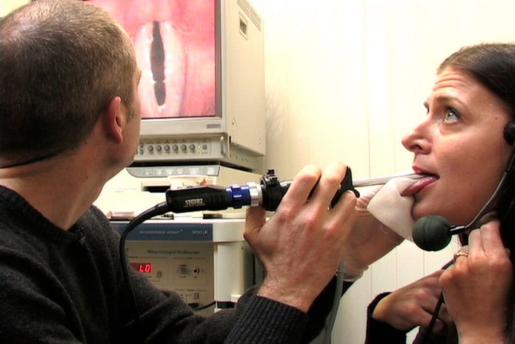
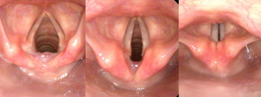
For hoarseness, we are generally interested in the vocal cords, located posterior to the thyroid cartilage. The vocal cords can easily be visualized from above, by passing a device into the pharynx. A rigid endoscope views the larynx from the back of the mouth. A flexible endoscope views the larynx from the back of the nose.

The Pharynx
The space inside the throat above the larynx is the pharynx. It is surrounded by muscles and the hyoid bone. Changing the shape and size of the pharynx alters resonance.
The Epiglottis and Arytenoids
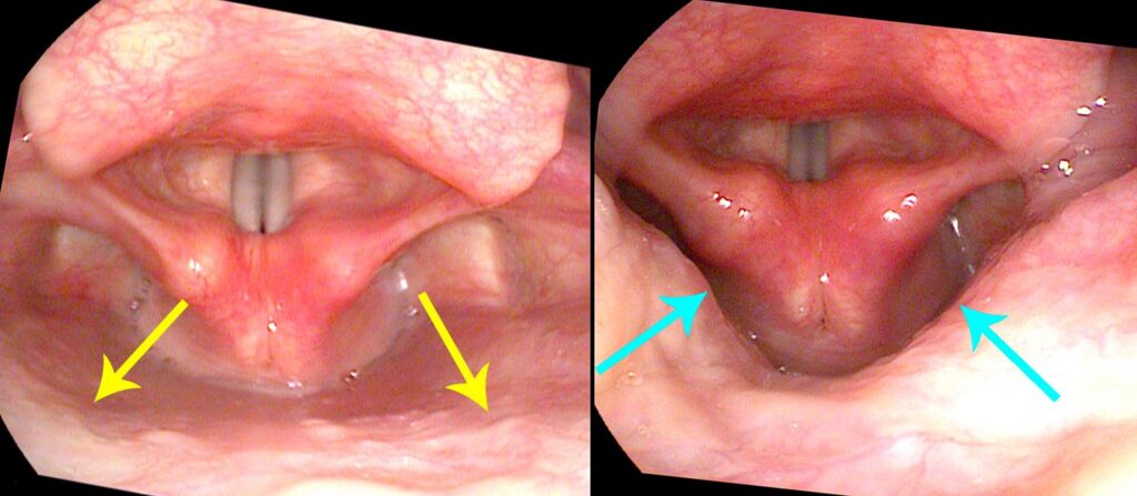
Technically the larynx has two cartilages that maintain its shape (thyroid and cricoid). A softer cartilage acts as a diverting valve during swallowing — the epiglottis. Two smaller cartilages open and close the vocal cords (arytenoids), and some miniscule cartilages sit on the arytenoids seeming to act as a dam to prevent residual liquids in the piriform sinuses from entering the airway.
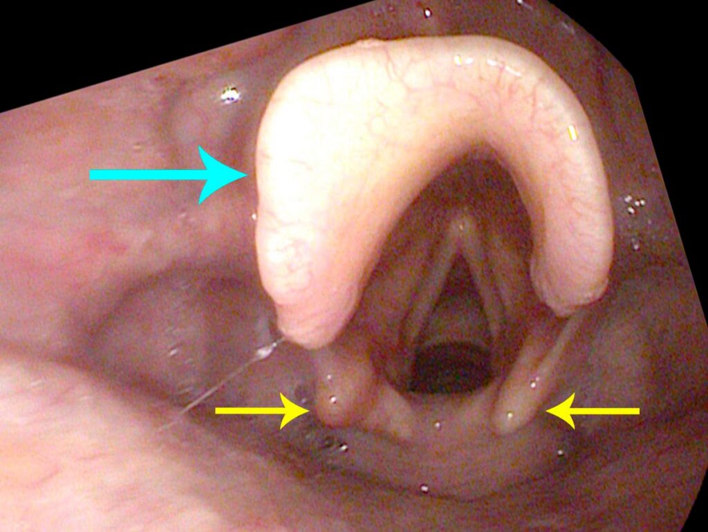
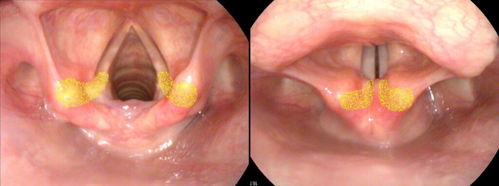
The front of the thyroid cartilage is triangular or tent-like in shape, suspending and protecting the vocal cords, with the Adam’s apple representing the apex. Inside, the airway is essentially a round tube with the vocal cords narrowing the airway, acting as a valve — technically, the glottis. The vocal cords narrow the opening to a triangle. During exhaling (breathing out), the vocal cords narrow the triangle to keep some back pressure in the lungs. During phonation, the vocal cords come almost completely together to form a narrow slit. Air passing between the vocal cords sets them vibrating and generates sound.
The machinery of the voice box is elegantly simple, each component serving a specific purpose.
The Intrinsic Muscles
There are 10 internal laryngeal muscles, vocal ligaments and some glands for lubrication. This is all covered with delicate, nearly translucent mucosa, tinted pink when viewed from far away by the blood flowing beneath it. All of this sounds complicated, but the machinery of the voice box is elegantly simple, with each component serving a specific purpose.
There are five muscles on each side of the larynx: TA (ThyroArytenoid), LCA (Lateral CricoArytenoid), PCA (Posterior CricoArytenoid), CT (CricoThyroid), and IA (InterArytenoid). With the various muscles changing the position, length and tension of the vocal cords, quite a range of sounds can be generated. Since each muscle is paired, any asymmetric contraction represents a probable weakness — something that becomes especially important in neurologic and muscular injuries.
What you learned
- The larynx is a valve with three jobs: regulate breathing, create sound, and keep food out of the lungs.
- Three cartilages maintain its shape — the thyroid and cricoid are structural; the epiglottis diverts food during swallowing.
- The arytenoids open and close the vocal cords; asymmetric arytenoid motion signals a potential neurologic problem.
- Ten intrinsic muscles in five paired groups control cord position, length, and tension — the full range of human voice.