The LCA muscle brings the vocal cords from an open breathing position together to a closed position in preparation for making sound. It is the primary adductor of the vocal cords.
The Lateral CricoArytenoid (LCA) Muscle
The LCA muscle brings the vocal cords from an open breathing position together to a closed position in preparation for making sound. This muscle is located parallel and just lateral to the TA muscle within the vocal cord. It is not particularly visible on an endoscopic exam although the effect of the contraction of the muscle is easily visualized.
The LCA muscle is attached to the outer end of the arytenoid cartilage, which acts like a lever. The other end of the arytenoid lever is the vocal process, a white cartilage visible towards the posterior of the membranous vocal cord. Contraction of the LCA muscle rotates the vocal process to the midline and if necessary beyond the midline.
When the LCA muscle on one side is injured, the opposite healthy LCA can develop enough strength to push its vocal process across the midline — a sign of compensation.
When relaxed, with the other muscles functioning normally, the vocal process is at the apex of an approximately 160°-angled, gently curving corner, formed by the membranous vocal cord and the soft tissue posterior to it. During normal contraction this corner disappears to form a straight line.
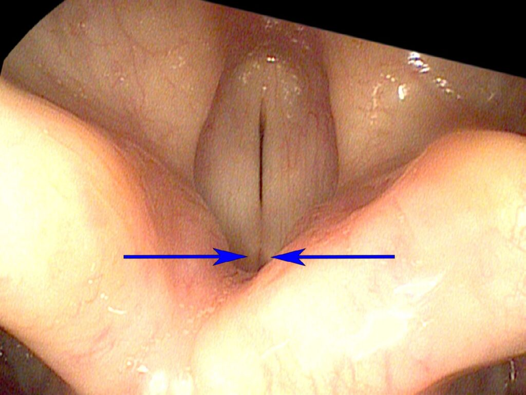
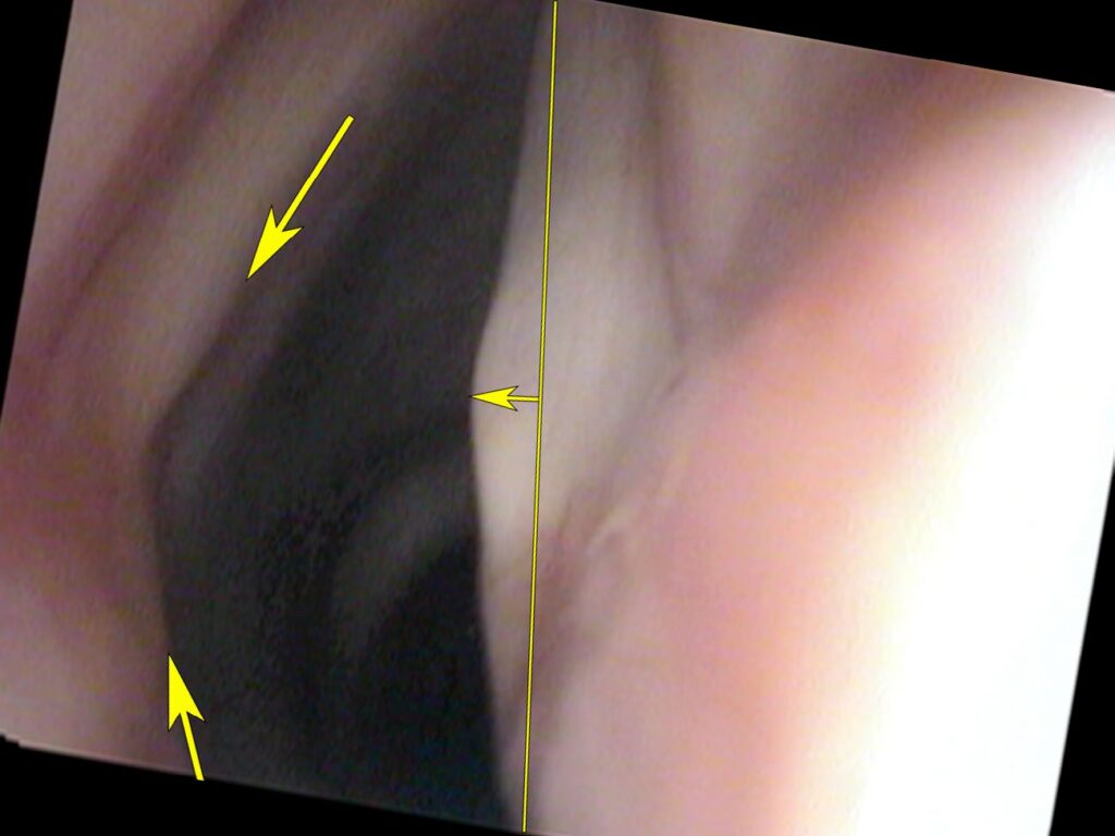
Compensation may occur when the LCA muscle on one side is injured. The opposite healthy LCA muscle has or can develop enough strength to move the vocal process across the midline and many times can reach or approach the vocal process on the weaker side. This may result in an inversion of the angle such that the vocal process now protrudes into the airway.
What You Learned
- The LCA is the primary adductor — it closes the vocal cords from the breathing position in preparation for phonation.
- It works via the arytenoid lever — pulling the outer end of the arytenoid rotates the vocal process to the midline.
- Crossing the midline signals compensation — a healthy LCA can push its vocal process past center to compensate for a weak contralateral LCA.
- Effect is easily visualized — though the muscle itself is not directly seen, the movement of the vocal process clearly reveals its action.