
Office endoscopy has reached the point where I consider the high-definition office exam the equivalent of an optical biopsy. Cancer and papilloma can be visually differentiated from benign lesions. In many cases, surgery is reserved for treatment — not diagnosis.
Optical Margins
The high-definition office endoscopic exam has reached a level of resolution where I consider it equivalent to an optical biopsy. Several consequences follow from this:
- Cancer and papilloma can be visually differentiated from benign lesions.
- Granulomas, polyps, smoker’s polyps, and nodules do not need a biopsy for diagnosis — surgery is reserved for treatment.
- All biopsies have the potential to be excisional (definitive treatment) rather than incisional (diagnostic only — and frequently non-diagnostic).
- Cancer margins are better defined with selective color imaging on endoscopes than with microscopes.
- There is no need to remove normal tissue to determine pathologic margins. This “assurance” by the pathologist is an illusion.
Case 1 — Polyps with Capillary Ectasias
A typical viewpoint for a vocal cord lesion from near the tip of the epiglottis, taken with an HD 720p chip camera, shows a reddish swelling on the left vocal cord. From this standard distance, a surgeon might not be certain whether to take this patient to surgery.
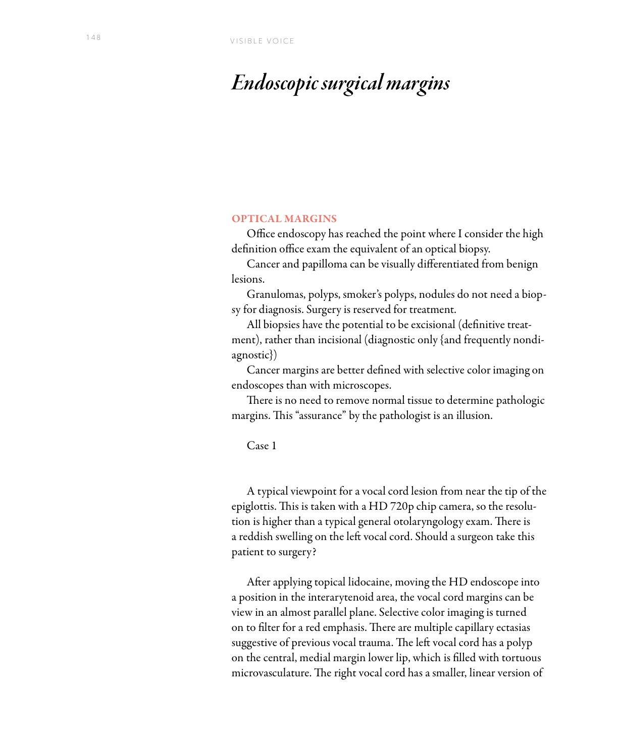
After applying topical lidocaine and moving the HD endoscope into a position in the interarytenoid area, the vocal cord margins can be viewed in an almost parallel plane. Selective color imaging is turned on to filter for red emphasis. There are multiple capillary ectasias suggestive of previous vocal trauma. The left vocal cord has a polyp on the central, medial margin lower lip, filled with tortuous microvasculature. The right vocal cord has a smaller, linear version of the same type of lesion.
The surgical microscopic view of the vocal cords at the time of surgery to remove the left and right polyps with capillary ectasias shows comparable resolution to the office view. They were removed to improve the voice — not for diagnostic purposes.
Cancer Surgery — What Matters Is What Currently Remains
In cancer surgery, what matters is what currently remains — not what the pathologist said after a surgery. There is no value in removing normal tissue as a margin. Removing normal tissue has a cost: it creates the illusion of confidence where there should be careful watching. Pathologic margins create confidence where there shouldn’t be.
Case 2 — Mapping Tumor Over Time
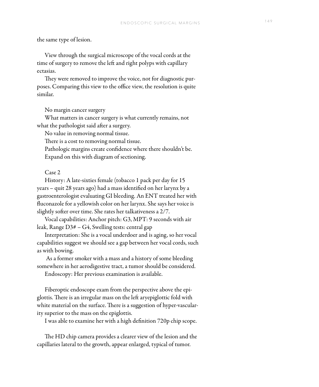
A late-sixties female — tobacco 1 pack per day for 15 years, quit 28 years ago — had a mass identified on her larynx by a gastroenterologist evaluating GI bleeding. An ENT treated her with fluconazole for a yellowish color on her larynx. She says her voice is slightly softer over time. She rates her talkativeness a 2/7.
Vocal capabilities: anchor pitch G3, MPT 9 seconds with air leak, range D3# – G4, swelling tests showing a central gap. Interpretation: she is a vocal underdoer and aging, so her vocal capabilities suggest a gap between her vocal cords such as with bowing. As a former smoker with a mass and a history of some bleeding, a tumor should be considered.
The HD chip camera provides a clearer view of the lesion and the capillaries lateral to the growth appear enlarged — typical of tumor. Selective color imaging highlights the hypervascularity surrounding the lesion, with capillary loops visible within the mass. This mass was squamous cell carcinoma. Nine months later, she has no symptoms.
One year later, her examination reveals vascular changes. Selective color imaging shows the left epiglottis covered with ectatic capillaries with large feeding vessels. Moving very close to the left epiglottis reveals a clear margin between normal vessels on one side and all of the capillary loops in the center of the photo — with feeding vessels on the periphery appearing very dilated. This tissue was peeled off both sides of the epiglottis and removed from the tongue base with a laser. At 2 years 6 months post initial surgery, a final island of abnormal capillaries was identified along a tissue ridge and treated in the office with a KTP laser.
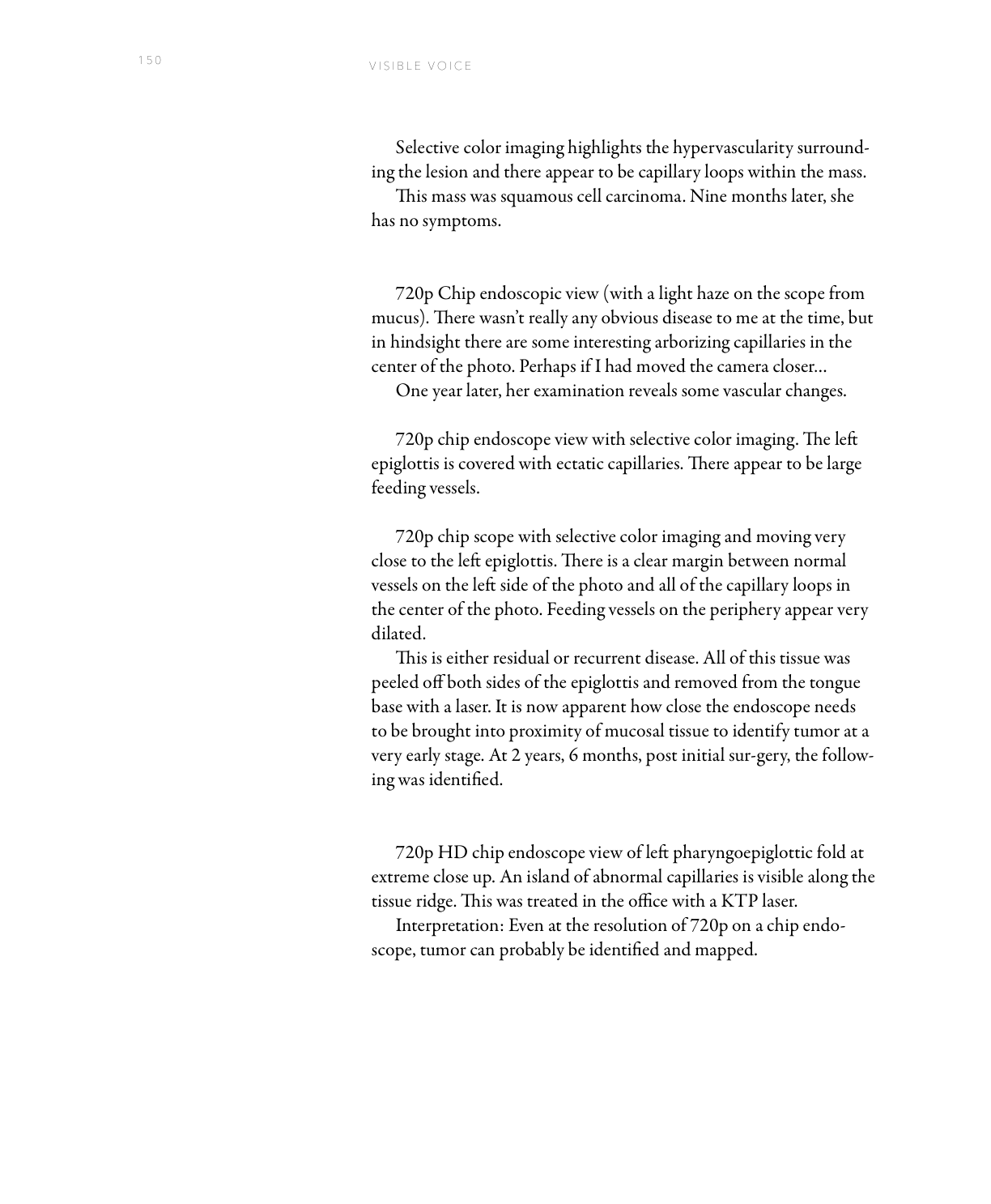
It is now apparent how close the endoscope needs to be brought into proximity of mucosal tissue to identify tumor at a very early stage. Even at the resolution of 720p on a chip endoscope, tumor can probably be identified and mapped.
What You Learned
- HD office endoscopy is an optical biopsy — benign lesions (granulomas, polyps, nodules) can be diagnosed visually; only treatment requires the operating room.
- Capillary architecture defines tumor margins — irregular, multilobulated, enlarged feeding vessels with perpendicular capillary loops distinguish malignancy from benign findings.
- Pathologic margins are an illusion of confidence — what matters in cancer surgery is what remains visible now, mapped with selective color imaging, not what the pathologist reported after a prior excision.
- Serial close-up exams map tumor evolution — comparing frame-by-frame images over months reveals recurrence or residual disease at an earlier, more treatable stage.