
Perspectives · Airway
When symptoms of cough and hoarseness might be hidden
How subglottic stenosis may hide for years behind a normal-looking larynx — and why understanding physics and airflow provides an accurate method for diagnosis.
By James P. Thomas, MD · Laryngology
Diane complains of a cough. It is chronic, occurs hundreds of times a day, mostly dry, but every so often productive, comes on hard and then eases off for a while. Her chest feels tight, too. She says she can feel her lungs. She thinks her breathing is okay, or at least she has been told that it is after testing by her lung doctor. When I probe specifically though, she admits that while she is golfing or active she does feel a constriction breathing in. She is also hoarse, with poor vocal endurance, which started almost a year ago, and she attributes it to allergies from her recent move here from the East. Her throat feels scratchy, too. She is in her late 50s.
A note on privacy: the patient below is a composite. Her name and a few identifying details have been changed, but the story — the years of wrong turns, the normal tests, the diagnosis hiding just below the vocal cords — is typical of nearly everyone who finally arrives in my office with this problem.
Let’s consider her symptoms:
- A dry cough that sometimes turns productive. It comes and goes. She coughs, by her own estimate, hundreds of times a day.
- A chest that “feels tight.” She says she can feel her lungs.
- Breathing that seems fine at rest — she’s been told repeatedly that it’s fine — but a distinct constriction when she breathes in while golfing or otherwise exerting herself.
- A little noise when she pushes herself physically.
- A scratchy throat and a voice that tires quickly and gets worse the more she uses it.
Here is the road she had already traveled before she reached me: an albuterol inhaler from her primary doctor (no benefit); an ENT who scoped her years earlier, described “a growth,” and sent her to speech therapy for vocal cord dysfunction; one “weird” session of therapy she abandoned; and a pulmonologist who ran full pulmonary function testing (normal), allergy testing (normal), and recommended a $235 asthma inhaler she didn’t buy.
By the time Diane sits down across from me, she has been labeled with reflux, asthma, and vocal cord dysfunction — treated for all three — and never improved. She believes her doctors who say she is resistant to treatment. Still, when something isn’t working, one of my top considerations is an erroneous diagnosis, not necessarily by commission, but by omission.
Why it hides
The vocal cords are often the star of the show — but can be the curtain
The subglottis is the short segment of airway just below the vocal cords, at the level of the cricoid — the only complete ring of cartilage in the entire airway. When you pass a flexible scope through the nose, the cords open and close in the foreground. Behind and below them, the subglottis sits in shadow. If the cords look healthy and move well, the natural instinct is to stop looking — and to blame something invisible, like silent reflux!
A brief glimpse of the subglottis offers a hint. Besides not finding any problem with the vocal cords, her symptoms suggest something lower, and careful observation during inspiration offers a tantalizing but incomplete view of the subglottis. In fact, most of the subglottis is not seen from above the level of the vocal cords.
Record the video and slow it down. The cords abduct, and for a few frames an abnormality shows itself — that glimpse is a key to open a door.
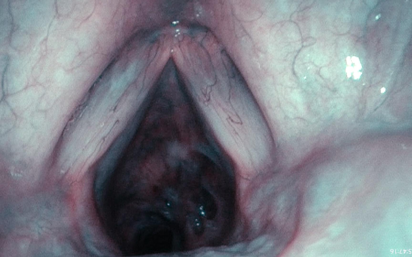
Don’t accept guessing. A little topical anesthesia — 4% lidocaine gargled on the larynx — then pass the endoscope through the vocal cords. Examine the airway all the way to the carina. The diagnosis stops being a hunch. This stenosis can explain all of her symptoms.
The endoscope even becomes a measuring tool for the stenosis. She takes a few breaths with the endoscope through the vocal cords, but above the stenosis. Then pass the endoscope (whose diameter we know) through the stenosis, have her keep breathing, and compare the effort. That gives you a sense of how much additional occlusion she could still tolerate.
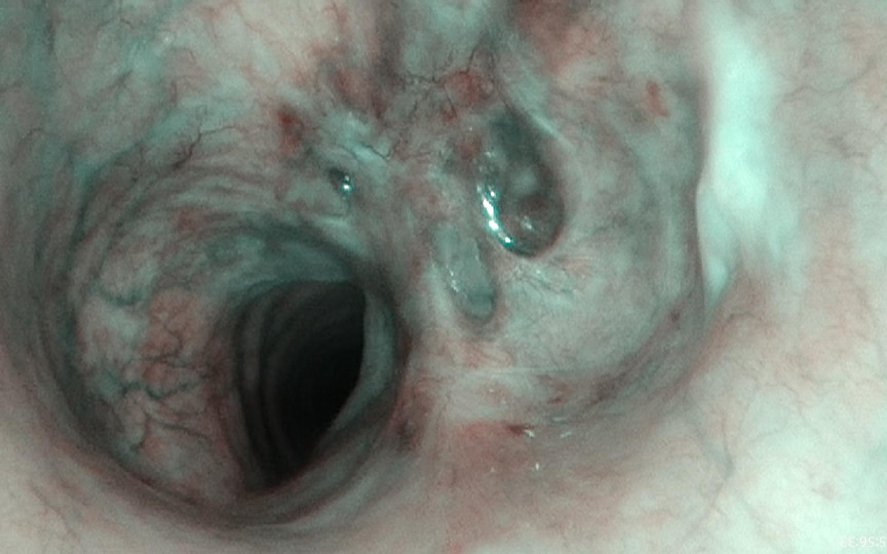
She actually is short of breath with activity — but had been convinced by her physicians that she wasn’t. So the stenosis explains the dyspnea. It is also the likely cause of the coughing: mucus collects on the edges of the stenosis, and she cannot generate enough force to clear the secretions. And because the cough never changes the size of the stenotic opening, her “tightness” is never relieved by it — she just needs another one. The stenosis even creates an artificially low subglottic pressure, so she drops easily into vocal fry without much effort.
So — one finding, a complete explanation, and nothing silent or invisible to invoke.
The physics everyone misses
Why the crisis is avoidable
This is the part I most want people — patients and physicians alike — to understand, because it explains both why the diagnosis is late and why it does not have to be. Airflow through a tube depends on the radius in a deeply unforgiving way: resistance rises with the fourth power of the radius. Halve the radius, and you don’t double the resistance — you multiply it roughly sixteen-fold.
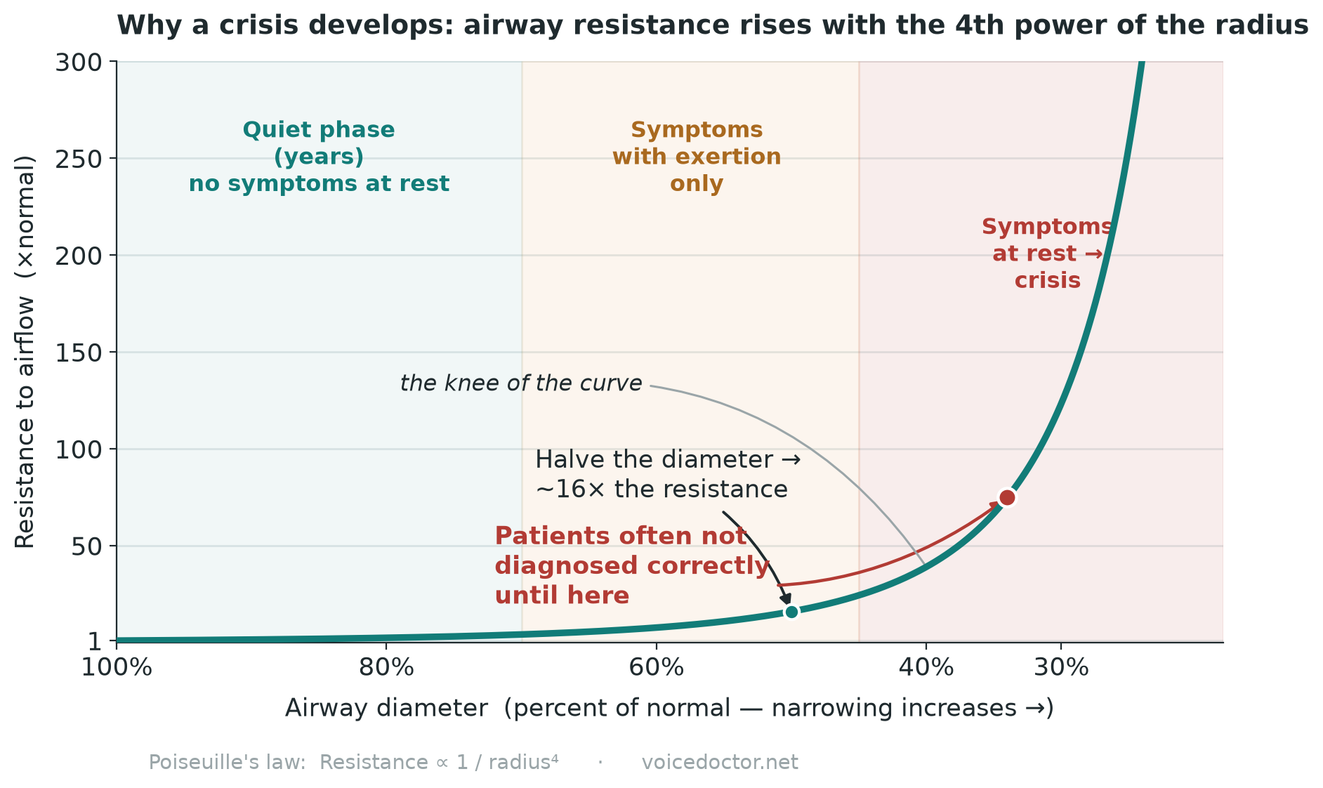
Picture what that does over years. Early on, resistance climbs but breathing at rest is easy — the patient notices only at peak exertion (Diane’s “constriction on the golf course”). This is the long, quiet, misdiagnosed phase.
Patients may accept explanations that aren’t working for a while, because a physician’s explanation carries weight. But they are not getting better — they are getting worse — so they hold two ideas at once: the doctors must be right, and there must be something else going on as well. It is that incongruency, not a crisis, that keeps Diane looking for an answer.
That is who arrives in my office. Not the emergency — by the time an airway reaches the steep part of the curve, that patient is getting a tracheostomy, not an appointment. The people who find me are still in the long middle, frustrated, and wanting to believe their doctors.
So my message to those doctors is the one on the door: listen carefully, and look closely. Listen to what she is telling you — and in Diane’s case, listen to the breathing itself. Then think about the physics, look below the cords, and you will see it.
Clinician note · what this breathing test could have said
Let me be honest about my own position first: I am a laryngologist, not a pulmonologist. I find this diagnosis by listening and by looking, not by reading flow loops. But her report is a useful teaching object, so here is what I understand it to show — and I would welcome correction from the people who read these all day.
What the report tabulated: FVC 3.32 L (94% predicted), FEV₁ 2.35 L (85%), FEV₁/FVC 71% (90%), FET 6.24 s, test quality graded A. Every one of those reads as mild and unremarkable.
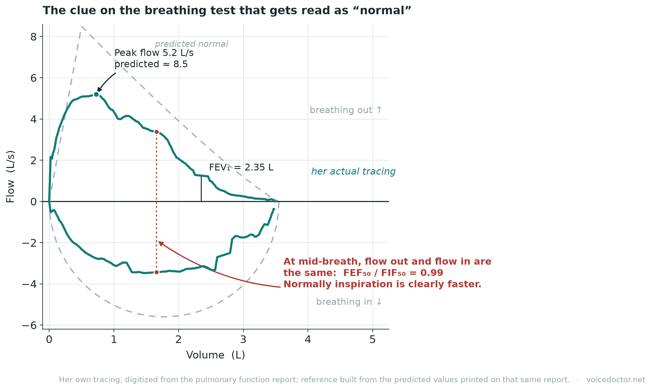
What it did not tabulate: PEF, FEF₅₀ and FIF₅₀ — the numbers built to interrogate the upper airway. Measured off her own tracing: peak expiratory flow about 5.2 L/s against a predicted near 8.5; FEF₅₀ ≈ 3.4 and FIF₅₀ ≈ 3.4.
The two indices built for this, and what they do here. FEF₅₀/FIF₅₀ — a ratio in the 0.85–1 band is the pattern of a fixed obstruction; at or above 1 the lesion is extrathoracic and variable; down near 0.3, variable and intrathoracic. Hers is 0.99, squarely in the fixed range. That is the one index on her whole study that lands. FEV₁/PEF, Empey’s index — above 10 mL per L/min makes central airway obstruction likely, and a more sensitive cut-off of 8 has been proposed. Hers is about 7.5. It does not flag.
And the caveat that matters most: spirometry is not a sensitive test for this. Measurable change in pulmonary function generally requires the lumen to be narrowed beyond roughly 80%, or the trachea to drop below about 8 mm. Diane’s airway was narrowed by a little more than half. So a nearly normal spirometry here was not a failure of the test, and not really a failure of the person reading it — it is what you should expect at her degree of narrowing. That is the resistance curve again, in a different costume: for years the numbers sit in range while the person steadily gets worse.
Which is the whole argument of this piece. The shape of the loop carries more than the table does, and the history carries more than either.
Two things worth carrying away. First, the quality grades on this report — the A for FEV₁ and FVC — describe the expiratory maneuver. A submaximal inspiration will conceal the very flattening you are hunting for, and nothing printed here would tell you it happened. Second, the asthma hypothesis had already been tested on this same report and had failed: FEV₁ went 2.35 → 2.59 L after bronchodilator, a change of 8.6% of predicted, short of the >10%-of-predicted threshold in the current ERS/ATS standard. She was treated for asthma anyway.
And when the breathing test reads “normal” but the patient is audibly turbulent on inspiration and limited on exertion, the upper airway deserves a direct look.
What can be done
A menu for treating the unknown
Subglottic stenosis is treatable in the sense that we can manage it, even if we cannot cure it. And the physics that made this so easy to miss now works in the patient’s favor: because resistance climbs with the fourth power of the radius, even a small gain in diameter buys a large drop in the effort of breathing. Smoothing a ridge in the airway helps in its own right — flow becomes more laminar, which means better breath support for a clearer voice, better clearance of secretions, and less breathlessness as the breathing quickens.
The operation I do divides the scar rather than removing it — excision invites recurrence — then stretches the airway with a balloon and injects steroid to slow the scar’s return. On its own that often settles into a yearly trip to the operating room. What changed things was Dr. Ramon Franco’s work on serial steroid injections: a quick office treatment as the breathing tightens again, quite often eliminating any further return to the operating room.
Operating room · first step
Stellate incisions
The scar is divided in a star pattern rather than excised, leaving a good deal of the mucosa intact. Cutting scar out invites it back.
Operating room · same sitting
Balloon dilation
The divided segment is stretched to restore diameter — the largest single gain of the sitting, and a small gain in radius is a large gain in airflow.
Operating room · same sitting
Steroid injection
Injected into the scar to slow its return before it starts.
In the office · awake
Serial steroid injections
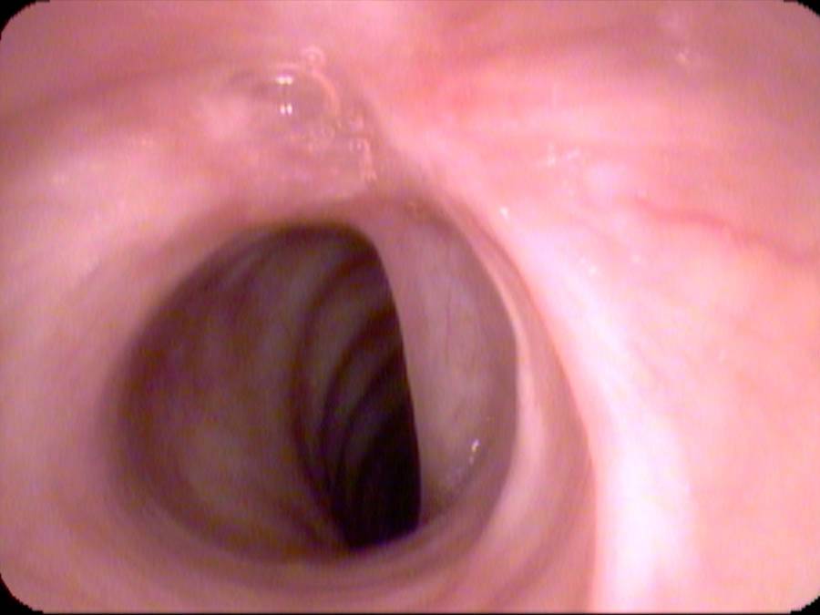
As the breathing tightens again — often four to twelve months later — an office injection shrinks the stenosis. Frequently this replaces the next trip to the operating room altogether.
Before
After
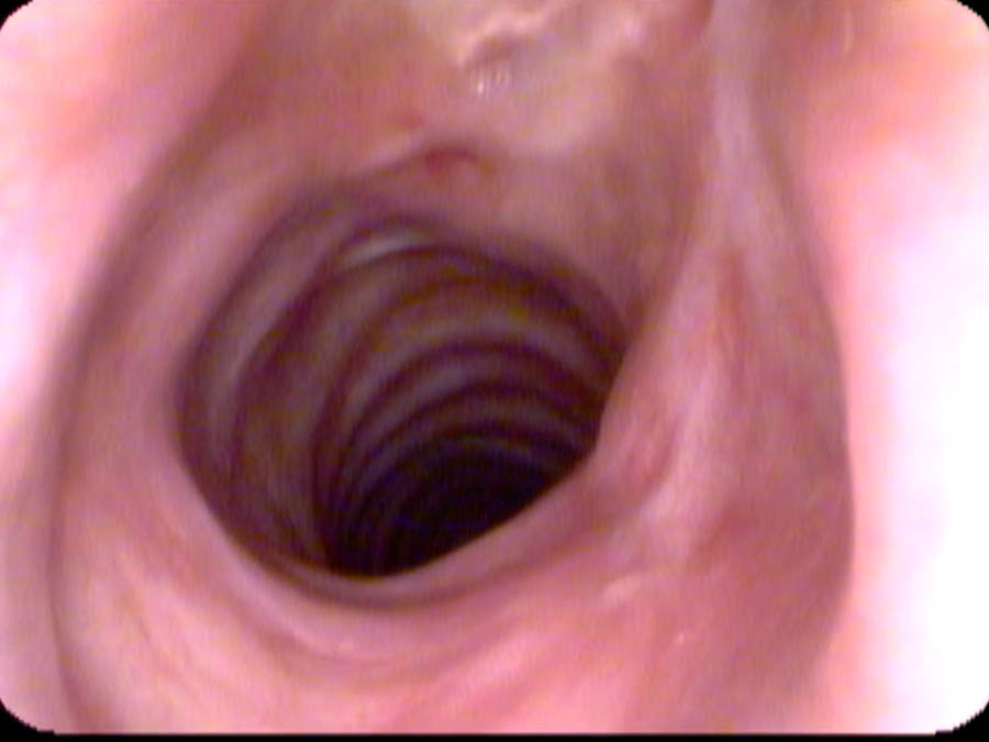
The surgical technique in detail — including what to avoid — is on Microlaryngoscopy: Stenosis. The office injection protocol is on Office Steroid Injection. The diagnostic method is on Below the Cords.
On cause
An honest counterweight to the textbook
Idiopathic simply means we don’t know, and that is honestly where we still stand.
What I was taught was more specific than that: autoimmune testing would often come back positive, the rheumatologist would treat what it showed, get the numbers under control, and the stenosis might improve along with them. I have not found that to be true. I still send patients to rheumatology — it is reasonable to look — but the testing comes back largely negative or equivocal. And even when ANA or ANCA is positive, bringing those numbers down has not, in my experience, changed the stenosis.
What I do see, nearly every time, is that the stenosis is hypervascular. Some of them bleed if you touch them. And steroid injected directly into the scar clearly works, at least to a degree. So even with the cause unknown, the scar does dissolve — and it is at least plausible that the steroid is treating a local inflammatory or autoimmune reaction rather than a systemic one. That fits what I see through the scope better than anything the blood tests have told me.
There is also a group studying and promoting an infectious cause. I will take that as a hypothesis — maybe it has a basis, maybe it explains some cases. But the presentation I heard was not persuasive, and low-grade chronic infection doesn’t usually resolve with antibiotics, so I remain skeptical.
Some cases are clearly post-intubation. Women have the disorder more often than men, though it occurs in both. For the rest, idiopathic remains the honest label. Someday we will figure it out. For now, the important thing is not to miss it — because we do have reasonable management strategies that give people a better quality of life, even if they are not curative.
When the voice complaint doesn’t explain the breathing complaint, keep going. The answer is often waiting just past the curtain.
James P. Thomas, MD
Sources & references
Subglottic stenosis overview — StatPearls, NCBI.
Spirometric indices in central airway obstruction (FEF₅₀/FIF₅₀, Empey’s FEV₁/PEF, and the narrowing required before spirometry changes) — Ampazis et al., J Clin Med 2024;13:6299 — doi:10.3390/jcm13216299.
Bronchodilator responsiveness criterion (>10% of predicted) — Stanojevic et al., ERS/ATS technical standard on interpretive strategies for routine lung function tests, Eur Respir J 2022;60:2101499 — doi:10.1183/13993003.01499-2021.
Degree of obstruction vs. airflow limitation (area vs. fourth-power nuance) — PMC5967982.
Monitoring adult subglottic stenosis with spirometry & dyspnea index — PMC9442627.
Awake serial intralesional steroid injections without surgery for idiopathic subglottic stenosis — Franco et al., The Laryngoscope 2018 — doi:10.1002/lary.26874.