
Cartilage: Giant Cell Tumor
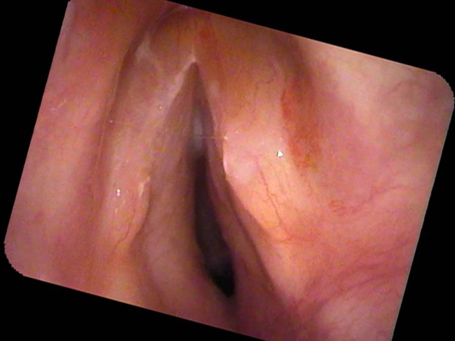
John Smith notes his voice has been worsening for the past three months, and is now losing it completely at times. Recently he has more difficulty breathing as well. “I feel like I’m breathing through a straw,” he complains. He smoked cigarettes for 30 years and he is finishing treatment for a tumor of his spine that seems to have completely gone away according to his oncologist’s most recent evaluation. Listening to his voice, it is deep pitched. He has almost no vocal range. He has almost no volume and runs out of breath after six seconds of making a sound. On endoscopic exam, the tissue beneath his vocal cords is squeezing in from the sides narrowing his airway.
Further testing with a CT scan shows that the foundation of his larynx, the cricoid cartilage, is almost completely replaced by tumor. A biopsy reveals a giant cell tumor of the cartilage.
Hemangioma
Hope Rouge, currently 50, recently had a surgery for snoring and was told by her ENT at that visit that her voice box looked abnormal. She came to see me. Although not a big concern to her, she says, “For about one year I have noticed some hoarseness. I sound like I have a cold all the time and my voice cuts out if I try to yell.” She smoked a pack of cigarettes a day for 25 years but she quit smoking seven years ago. Her voice is deep sounding, almost masculine in quality. When I test her voice, sound starts cutting out from about middle C upward. It sounds like something is touching her vocal cords.
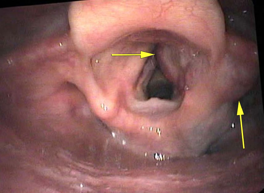
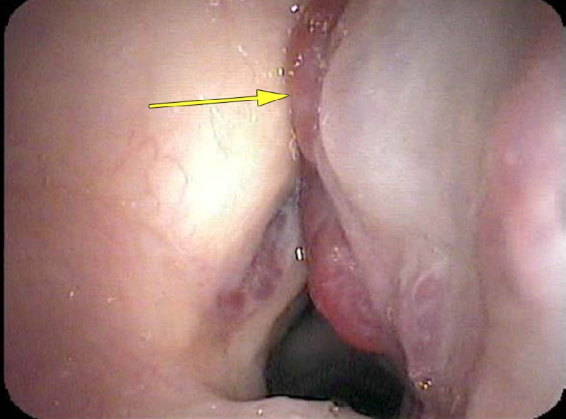
On endoscopic exam the edges of her vocal cords are normal. However, she has deep red, dilated blood vessels filling and enlarging the right false vocal cord. There are also a few on the left side. When she tries to make a sound, this hemangioma presses against the true vocal cords and stops the vibrations.
Five years after a surgical excision, she still had small remaining blood vessels on the right side but no further symptoms. She will continue to be examined with an endoscope on a regular basis or if any laryngeal symptoms return. Additionally, since her treatment, a new medical approach has been discovered where some blood vessel tumors will shrink with medical treatment with a beta blocker.
Although the mucosa is the most common cell type to become cancerous, any cell type in the larynx can become malignant — including nerve cells, blood vessel walls, fibrous cells, muscle cells, cartilage cells, or blood vessels.
What you learned
- Any cell type in the larynx — cartilage, blood vessels, nerve cells, fibrous tissue — can become malignant; hoarseness and airway narrowing may be the first sign.
- A giant cell tumor of the cricoid cartilage replaces the structural foundation of the larynx, narrowing the airway from below the vocal cords rather than affecting the cord surface directly.
- A hemangioma in the false vocal cord causes hoarseness by pressing down on the true vocal cords during phonation and dampening their vibrations.
- Some vascular tumors (hemangiomas) respond to beta-blocker medication, offering a non-surgical treatment option for selected patients.
- When a vocal cord cannot be visualized because of a mass in the false cord, the true cord pathology may be missed entirely — a reminder to look carefully above the true cords as well.