
The terms “vocal cord” and “vocal fold” are used interchangeably. Viewed from above they appear as two white bands; in cross-section they are more like a wedge — a muscle under a ligament, covered by mucosa.
Vocal Cords, Vocal Folds
What we term “vocal cord” is often compared to the edge of a string when viewed from above during vibration, and this is possibly where the term cord came from. Viewed in cross section though, the vocal cord doesn’t look like a cord at all. It is more of a wedge in shape. Still, musically it does function like a cord and many analogies to a cord or string, such as a comparison to the string on a guitar, can be made.
Some people call them “vocal folds,” which is a more apt description of their three dimensional visual appearance. They are each a fold of soft tissue rising from the edges of the airway. The terms vocal cord and vocal fold can be, and are, used interchangeably.
The mucosa is like a layer of silk draped over the edge of the ligament — and the mucosa vibrates when air passes rapidly by it.
The vocal cord is essentially a muscle under a ligament with mucosa covering both of them. A layer of lubricant, the lamina propria, lies between the mucosa and the ligament-muscle combination. The muscle tightens and loosens to change the pitch of the vibrations. The lubricating layer allows the mucosa on the surface to vibrate easily. The muscle may oscillate to a small degree, like a string on a guitar or piano, but the lining on the surface is the primary oscillator or generator of sound.
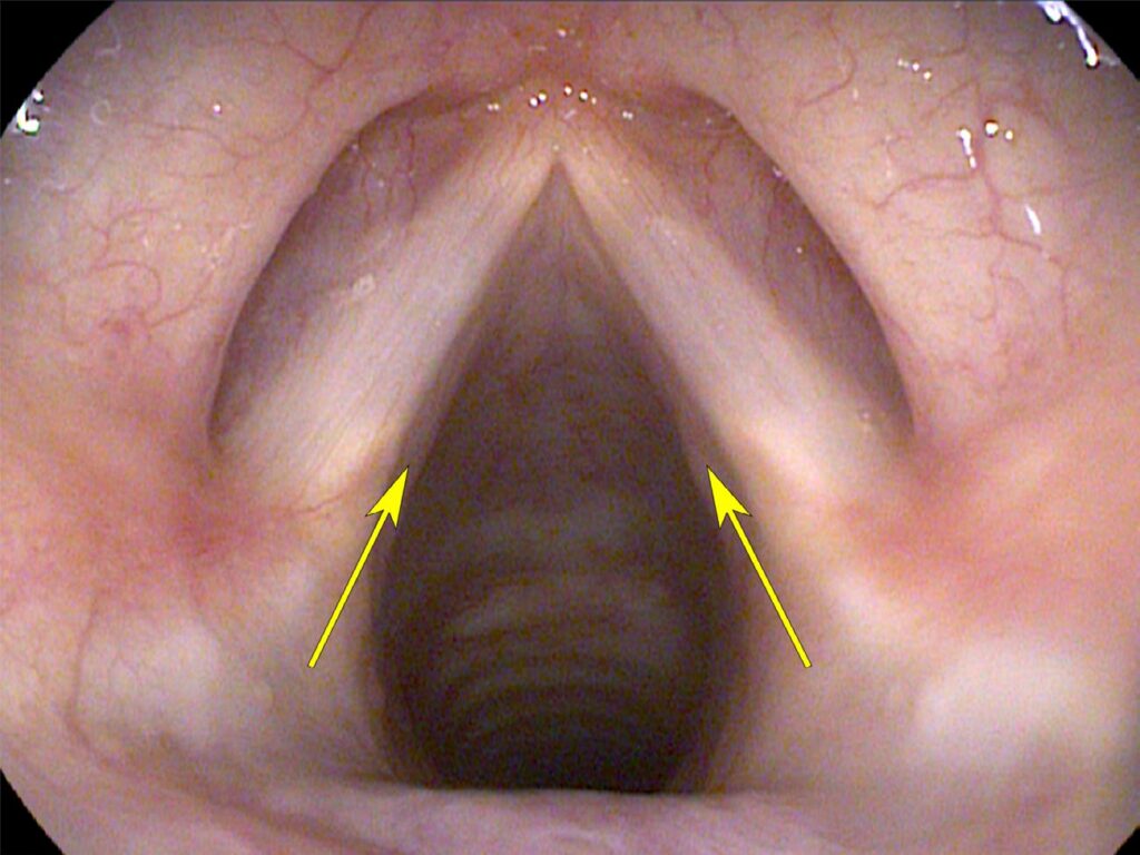
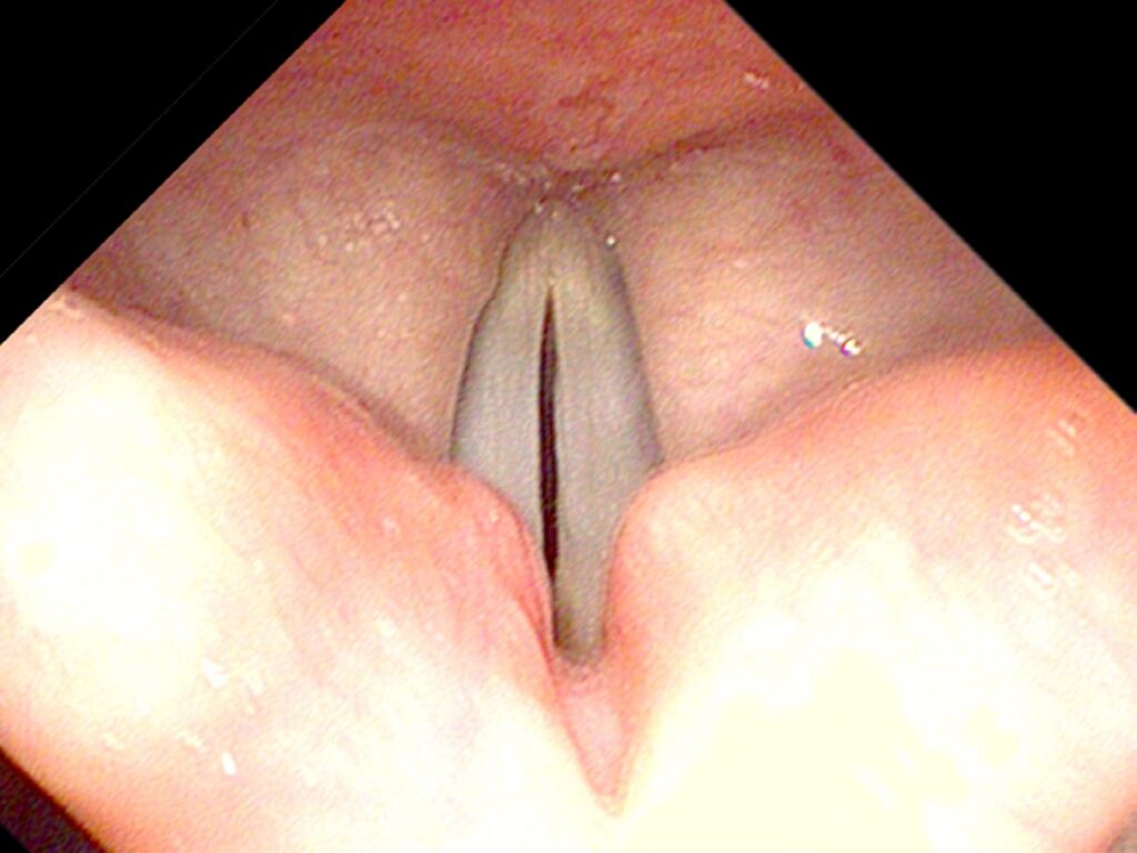
The vocal cords have two general positions: ABducted and ADducted. When ABducted, they are apart and in the configuration of a V — the breathing position. When ADducted the vocal cords should come essentially into near alignment, often almost parallel to each other. The lungs generate pressure below and air can then be passed through the vocal cords from the windpipe below. As air passes through this narrow slot between the vocal cords, the mucosa starts to vibrate, creating sound.
What You Learned
- “Vocal cord” and “vocal fold” mean the same thing — the terms are interchangeable; “fold” better describes the three-dimensional shape.
- Layers matter — the cord is a muscle beneath a ligament beneath mucosa, with the lamina propria lubricating the interface.
- The mucosa is the primary vibrator — it acts like a layer of silk draped over the ligament edge, oscillating as air passes through.
- Two positions: ABducted and ADducted — ABducted (apart, V-shape) is for breathing; ADducted (together) is for phonation.